Male Pelvic Floor Muscle Ultrasound: How It Works Without an Internal Exam
When most people think about a pelvic floor assessment, they picture an internal exam. While that's one way to evaluate the pelvic floor, it's certainly not the only option.
Rehabilitative pelvic floor muscle ultrasound gives us another way to assess how your pelvic floor muscles are working without performing an internal examination. Using ultrasound, we can watch the muscles move in real time, evaluate bladder support and coordination, and help you learn how to contract and relax your pelvic floor more effectively.
For many people, this technology makes pelvic floor therapy feel less intimidating while providing valuable information that helps guide treatment.
Let's take a closer look at how pelvic floor ultrasound works, what it shows, and why it's becoming such a valuable tool in pelvic floor health.
What Is Pelvic Floor Muscle Ultrasound?
Pelvic floor muscle ultrasound is a rehabilitative imaging technique that allows pelvic health therapists to observe pelvic floor structures and muscle function in real time.
Unlike diagnostic ultrasound, which is used to identify injuries or disease, rehabilitative ultrasound is used to assess movement, coordination, muscle activation, and motor control.
It provides both the therapist and the patient with immediate visual feedback on what's happening during pelvic floor contraction, relaxation, and bearing down. We can even see the bulb of the penis and muscle contraction around it too!
One of the biggest advantages is that it can often be performed without an internal examination.
Who Can Benefit from Pelvic Floor Ultrasound?
Pelvic floor ultrasound can be helpful for a wide variety of people, including those experiencing:
Urinary urgency
Frequent urination
Bladder leaks, bladder incontinence
Fecal incontinence
Constipation
Pelvic pain
Anal pain
Erectile dysfunction
Premature ejaculation
Ejaculatory pain
Pelvic floor muscle dysfunction
Difficulty relaxing the pelvic floor
Difficulty coordinating pelvic floor contractions
Recovery after any pelvic cancer treatment like prostate or rectal cancers
It's also a great option for anyone who isn't comfortable with an internal pelvic floor assessment or prefers to start without an internal exam.
While ultrasound doesn't replace an internal examination, it provides a wealth of useful information to guide rehabilitation with or without the internal examination. Two Different Ultrasound Approaches
There are two common ultrasound views used during pelvic floor rehabilitation.
Two Different Ultrasound Approaches
There are two common ultrasound views used during pelvic floor rehabilitation.
Transabdominal Ultrasound
During a transabdominal assessment, the ultrasound probe is placed above the pubic bone.
Here we can visualize:
Bladder capacity when full
Your ability to empty your bladder
Pelvic floor muscle activity viewing bladder base movement lying down and standing
To get the best image, the bladder should be pretty full before the scan. We generally ask folks to drink lots of fluid (300-600ml) and postpone voiding for at least 60-90 minutes prior to the scan.
What Can We Learn from the Bladder View?
Watching the bladder move provides important information about pelvic floor function.
When someone attempts to contract their pelvic floor muscles, we would expect to see the bladder base lift symmetrically upward.
If little movement occurs, it could suggest a few things:
Pelvic floor muscle weakness
Poor muscle coordination
Pushing instead of contracting
Increased muscle tension
Fascial or connective tissue deficits
Difficulty performing the contraction correctly
Measuring Bladder Volume
Another advantage of transabdominal ultrasound is bladder volume measurement.
This can help evaluate:
Post-void residual urine
Urinary retention
Functional bladder capacity
Bladder filling patterns
Urinary urgency and frequency
Understanding how full the bladder actually is can provide valuable context when someone reports feeling a strong urge to urinate.
Sometimes the bladder is actually full and the urge is proportionate to bladder fullness. Other times, the sensation of urgency occurs even when very little urine is present.
The visual confirmation helps shape treatment decisions and patient perception of bladder urge.
Transperineal Ultrasound
The second pelvic floor muscle ultrasound view is the transperineal view.
Instead of placing the probe over the lower abdomen, it's positioned underneath the scrotum above the anus.
This produces a “side-profile” view of the pelvic floor from front to back, like what is seen in this image below:
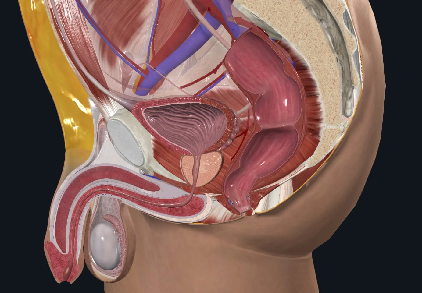
Image: Complete Anatomy
From this perspective, we can see the following structures:
Pubic symphysis
Bladder
Urethra
Striated urethral sphincter
Bulb of the penis
Bulbocavernosus muscle
Bladder neck
Rectum
Anorectal junction
Anal sphincter muscles
Levator ani muscles (pelvic floor muscles)
Seeing these structures move together provides valuable insight into how well the pelvic floor is functioning.
Why Function Matters More Than Numbers
One of the most important things to understand about male pelvic floor ultrasound is that there currently isn't a well-established set of normal values.
We can identify and observe movement of structures like:
The bulbocavernosus muscle
The urethral sphincter
The anorectal junction
The penile base
Levator ani muscles (pelvic floor contraction, relaxation, and bearing down)
Most of the available research does not indicate a universally adopted set of normal values yet, and because of that, pelvic health therapists generally focus less on exact measurements and more on how the pelvic floor moves. If you’re an actual researcher, measurements matter more for the study rather than real-life clinical application. Movement quality, patient feedback, often tells us much more than a measurement at this point.
What Does Pelvic Floor Movement Look Like?
During a pelvic floor contraction, we generally expect to see the pelvic floor lift structures like the bulb of the penis, bladder, anorectal junction and urethra up toward the head.
During bearing down or a Valsalva maneuver, we expect the pelvic floor to descend.
We're also looking for:
Smooth coordination
Symmetrical movement
Complete relaxation after contraction
Appropriate timing
No contraction when we except relaxation or lengthening
These movement patterns help us determine whether the muscles are working together efficiently.
Common Dysfunction Patterns
Ultrasound can reveal several movement patterns that may contribute to symptoms.
These include:
Reduced Pelvic Floor Muscle Excursion
Sometimes the muscles don't move very much during contraction.
This may reflect muscle weakness, increased resting tension, restricted movement, or poor motor control.
Excessive Descent
In some people, the pelvic floor or organs descend a lot more than expected during bearing down.
This may suggest reduced pelvic tissue support or weakness but not always.
Dyssynergia
Dyssynergia occurs when muscles contract instead of relaxing during activities like bearing down, attempting a bowel movement, or when you’re trying to empty your bladder.
This paradoxical contraction can contribute to constipation, incomplete bladder or bowel emptying, hemorrhoids, anismus, or pelvic pain and tightness.
Delayed Relaxation
Some people can contract their pelvic floor easily but struggle to let the muscles relax afterward.
Persistent muscle tension may contribute to symptoms like pelvic pain, ejaculatory pain, anismus, urinary or bowel symptoms, or difficulty emptying the bladder and bowels.
Asymmetry
Sometimes one side of the pelvic floor moves differently than the other.
This lack of coordination may indicate muscle imbalance or altered motor control that can be addressed during therapy.
Why Visual Feedback Is Helpful
One of the biggest benefits of rehabilitative ultrasound is that patients can actually watch their muscles move for greater brain-body training.
Instead of trying to imagine what a pelvic floor contraction should feel like, you can see it happening on the screen.
That visual feedback helps your brain connect what you're feeling with what's occurring inside your body.
For many people, this improves body awareness and makes pelvic floor exercises much easier to learn.
Is Pelvic Floor Ultrasound a Replacement for an Internal Exam?
Not necessarily.
An internal pelvic floor examination still provides information that ultrasound cannot, such as direct assessment of muscle tenderness, tissue mobility and tone, and individual muscle palpation.
However, pelvic floor ultrasound can answer many important questions without requiring an internal examination.
For some patients, that's enough information to begin treatment.
For others, ultrasound becomes the first step before deciding whether additional examination is needed later or a second diagnostic tool to confirm palpation exam findings.
A Valuable Tool for Pelvic Floor Rehabilitation
Pelvic floor ultrasound has become one of my favorite tools because it helps bridge the gap between what patients feel and what's actually happening inside their bodies.
Whether someone is recovering after prostate cancer treatment, managing chronic pelvic pain, experiencing bladder leaks, or struggling with pelvic floor coordination, ultrasound provides real-time information that can guide treatment and improve exercise performance.
It's non-invasive, educational, and empowering for patients who want to better understand how their pelvic floor works.
This information is not intended to substitute for professional medical advice, diagnosis, or treatment. Always seek the advice of a healthcare professional with any questions you may have regarding treatment, medications/supplements, or any medical diagnoses. This information is intended for educational purposes only and is in no way to substitute the advice of a licensed healthcare professional.